Thyroid Ultrasound Explained: How Doctors Spot Nodules and Assess Cancer Risk
Most people don't think about their thyroid until a doctor mentions a "nodule" during a routine checkup. It’s a scary word that immediately raises questions: Is it cancer? Do I need surgery? Will this change my life?
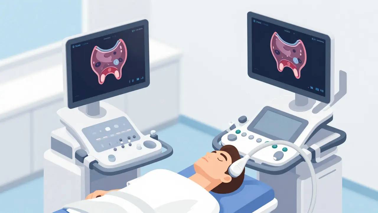
The good news is that the vast majority of thyroid nodules are benign. But knowing which ones are harmless and which ones need closer attention requires more than just feeling your neck. This is where thyroid ultrasound comes in. It is the gold standard imaging test for evaluating thyroid health, using sound waves to create detailed pictures of the gland without any radiation exposure. Unlike a physical exam, which can only detect large lumps, ultrasound picks up tiny abnormalities that might otherwise go unnoticed.
If you’ve been told you have a thyroid nodule, understanding how ultrasound works-and what those technical terms on your report actually mean-can take the fear out of the process. Let’s break down exactly how doctors use this technology to assess your risk and decide on the next steps.
Why Ultrasound Is the First Choice for Thyroid Imaging
You might wonder why your doctor didn’t order an MRI or a CT scan. While those tests are great for looking deep inside the body, they aren’t the best tools for the thyroid. The thyroid sits right under your skin in the front of your neck, making it perfect for ultrasound technology.
Ultrasound uses high-frequency sound waves (between 7.5 and 18 MHz) to bounce off tissues and create images. Because the thyroid is superficial, these high frequencies provide incredibly sharp resolution. In fact, studies show that ultrasound detects nodules in 19% to 68% of people, whereas physical palpation by a doctor only finds them in 2% to 21% of cases. That’s a massive difference in detection capability.
Here is why ultrasound beats other methods:
- No Radiation: Unlike CT scans or nuclear medicine scans, ultrasound uses no ionizing radiation. This makes it safe for repeated follow-ups over years.
- Real-Time Detail: It shows blood flow (using Doppler technology) and tissue texture instantly.
- Cost-Effective: In the US, a procedure typically costs between $200 and $500, far less than advanced cross-sectional imaging.
- Better Sensitivity: Ultrasound catches small nodules that CT and MRI often miss entirely.
Nuclear scans can tell if a nodule is "hot" (overactive) or "cold" (underactive), but they can’t tell you if it’s cancerous. Only ultrasound provides the structural details needed to judge malignancy risk accurately.
Decoding Your Report: The TI-RADS System
When you get your ultrasound results, you won’t just see a picture. You’ll see a score. This is likely based on the ACR TI-RADS system, which stands for Thyroid Imaging Reporting and Data System, a standardized scoring method created by the American College of Radiology to categorize nodule risk. Think of it as a traffic light system for your thyroid nodule.
Radiologists look at five specific features of each nodule to assign points:
- Composition: Is it solid, fluid-filled (cystic), or mixed?
- Echogenicity: How dark or bright does it look compared to surrounding tissue? Darker (hypoechoic) areas can be more suspicious.
- Shape: Is it wider than it is tall, or taller than it is wide? A "taller-than-wide" shape is a red flag.
- Margins: Are the edges smooth and clear, or jagged and ill-defined?
- Echogenic Foci: Are there tiny bright spots inside? These microcalcifications can indicate calcium deposits associated with cancer cells.
Each feature gets a point value. The total score places the nodule into a category from TR1 to TR5:
| Category | Points | Risk Level | Cancer Probability |
|---|---|---|---|
| TR1 | 0 | Benign | <0.3% |
| TR2 | 2 | Not Suspicious | <1.5% |
| TR3 | 3 | Mildly Suspicious | <4.8% |
| TR4 | 4-6 | Moderately Suspicious | <9.1% |
| TR5 | 7+ | Highly Suspicious | <35% |
This system helps doctors avoid unnecessary biopsies for low-risk nodules while ensuring high-risk ones get immediate attention.
When Do You Need a Biopsy?
An ultrasound doesn’t diagnose cancer; it estimates risk. To know for sure, doctors often perform a Fine-Needle Aspiration (FNA) biopsy. This involves using a thin needle to withdraw cells from the nodule for lab analysis. Ultrasound guides the needle with precision, reducing failed samples from 25% to less than 5%.
So, when does a nodule get biopsied? It depends on both its size and its TI-RADS score. Here are the general guidelines:
- TR1 & TR2: Usually no biopsy needed unless the nodule is very large and causing symptoms like swallowing difficulties.
- TR3: Biopsy recommended if the nodule is larger than 2.5 cm.
- TR4: Biopsy recommended if the nodule is larger than 1.5 cm.
- TR5: Biopsy recommended if the nodule is larger than 1 cm.
Nodules smaller than 5 mm are rarely biopsied because even if they were malignant, they are unlikely to cause harm. For many patients, especially those with small, low-risk nodules, "active surveillance"-just watching the nodule with regular ultrasounds-is the safest and most effective approach.
What If the Results Are Indeterminate?
About 15% to 30% of biopsies come back as "indeterminate." This means the cells look weird, but not definitively cancerous. This used to lead to a lot of unnecessary surgeries. Today, we have better options.
Molecular testing has changed the game. By analyzing the DNA of the nodule cells, doctors can predict whether an indeterminate nodule is likely to behave aggressively. This has reduced unnecessary thyroidectomies by nearly 50%. Even with molecular testing, however, you will still need periodic ultrasounds to monitor the nodule’s growth.
Recent advances in artificial intelligence are also helping here. A 2023 study published in *Nature Scientific Reports* showed that AI models could achieve 94.2% diagnostic accuracy by analyzing nodule shapes and patterns, outperforming traditional methods. While AI isn’t replacing radiologists yet, it’s becoming a powerful second opinion tool.
Preparing for Your Ultrasound
The good news? There’s almost nothing you need to do to prepare. Unlike abdominal ultrasounds, you don’t need to fast or drink water beforehand. Just wear comfortable clothing that allows easy access to your neck area.
During the exam, a technician will apply gel to your neck and move a transducer (the handheld device) across your skin. You might feel slight pressure, but it shouldn’t hurt. The whole process takes about 15 to 20 minutes. They will also check your cervical lymph nodes, as thyroid cancer can sometimes spread there first. Don’t skip this part-it’s crucial for accurate staging.
Common Questions About Thyroid Ultrasounds
Is a thyroid ultrasound painful?
No, the ultrasound itself is painless. You may feel some mild pressure as the technician moves the probe over your neck. If a biopsy is performed afterward, you might feel a brief sting from the needle, similar to a blood draw.
How often should I get a thyroid ultrasound if I have nodules?
For low-risk nodules (TR1/TR2), doctors often recommend repeating the ultrasound every 2-3 years to check for growth. Higher-risk nodules may require annual monitoring. Always follow your specialist’s specific schedule.
Can ultrasound detect thyroid cancer early?
Yes, ultrasound is highly sensitive and can detect nodules as small as a few millimeters. However, detecting a tiny nodule doesn’t always mean it needs treatment. Many small cancers grow so slowly that active surveillance is safer than immediate surgery.
What does "hypoechoic" mean on my report?
Hypoechoic means the nodule appears darker than the surrounding thyroid tissue on the ultrasound image. While many hypoechoic nodules are benign, markedly hypoechoic nodules carry a slightly higher risk of malignancy and are weighted more heavily in the TI-RADS scoring system.
Do I need to stop taking thyroid medication before the test?
Generally, no. Thyroid medication does not interfere with ultrasound imaging. Continue taking your prescribed medications unless your doctor instructs otherwise.